Lowell S Su, MD, Christian W Ertl, MD FACS FACCWS, Leandra H Burke, BS. Michigan State University / Kalamazoo Center for Medical Studies
Introduction:
Calculous biliary disease represents the most common cause of gastrointestinal illness requiring hospitalization in young, healthy individuals. Approximately two-thirds of symptomatic patients present with multiple attacks, suggesting ongoing inflammation, chronic obstruction, and histological changes within the gallbladder wall. Here we present a rare case of transmural fibrotic gallbladder in a patient with no known previous gallbladder disease.
Case Report:
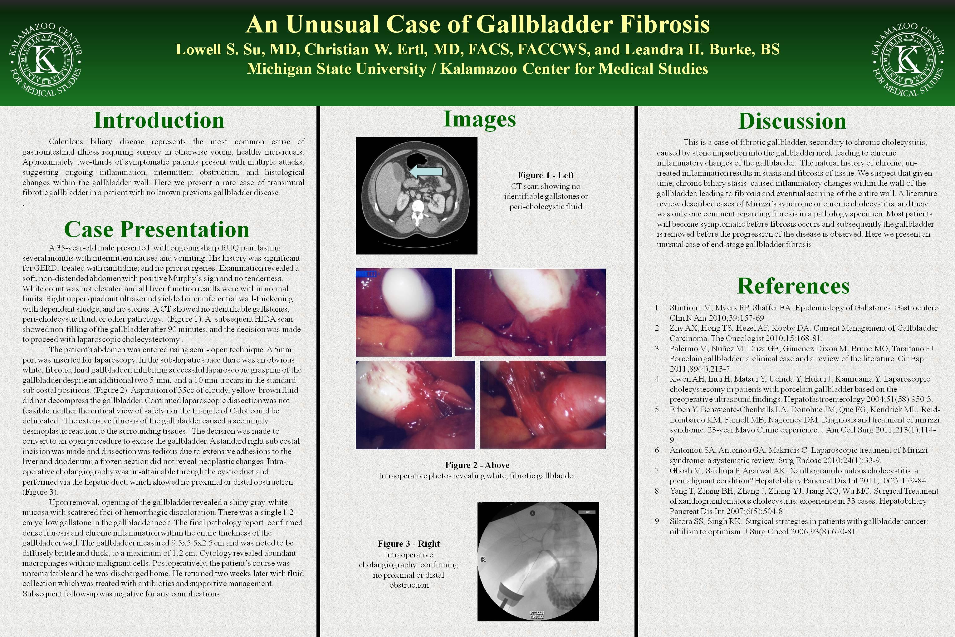
A 35-year-old male presented to our care with ongoing sharp RUQ pain lasting several months with intermittent nausea and vomiting. The patient had a history significant for GERD for which he took Ranitidine for symptom relief. The patient had no previous surgical history. Examination revealed a soft, non-distended abdomen with positive Murphy’s sign and mild epigastric tenderness. WBCs were not elevated and all liver function results were within normal limits. Abdominal ultrasound yielded circumferential wall-thickening of the gallbladder with dependent sludge, and CT showed no identifiable gallstone or pericholestic fluid. A HIDA scan showed non-filling of the gallbladder after 90 minutes, and a decision was made to proceed with elective laparoscopic removal of the gallbladder.
The patient’s abdomen was entered using standard Hassan technique. A 10- mm port was inserted for laparoscopy, followed by three 5-mm trocars in the standard sub costal positions. The abdomen was noted to have a white, fibrotic, hard gallbladder which prevented successful laparoscopic grasping of the gallbladder wall. Aspiration of 35 cc of cloudy, yellow-brown fluid was achieved and sent to pathology. An attempt to continue with laparoscopic dissection proved to be infeasible due to a difficult dissection, obliteration of the triangle of Calot, and a very fibrotic gallbladder with extensive inflammatory reaction and fibrinous adhesions to the peripheral tissues. The decision was made to convert to an open procedure to excise the gallbladder.
The final pathology report found dense fibrosis and chronic inflammation within the entire thickness of the gallbladder wall. The gallbladder measured 9.5 x 5.5. x 2.5 cm and was noted to be diffusely brittle and thick, to a maximum of 1.2 cm. Opening of the gallbladder revealed a shiny gray-white mucosal aspect with scattered foci of hemorrhagic discoloration. There was a single 1.2 cm yellow gallstone found impacted in the gallbladder neck. Cytology revealed abundant macrophages with no malignant cells. Postoperatively, the patient’s course was unremarkable and he was discharged home. He returned two weeks later with fluid collection which was treated with antibiotics and supportive management. Subsequent follow-up was negative for any complications.
Discussion:
We postulate that this is a case of chronic cholecystitis caused directly by stone impaction into the gallbladder neck leading to chronic inflammatory changes of the gallbladder which resulted in the stated pathology. Given time, chronic bile stasis results in inflammatory changes within the wall of the gallbladder, and fibrosis and scarring will eventually affect the entire wall. Most patients will become symptomatic before fibrosis occurs and consequently, the gallbladder is removed before the progression of the disease is observed. Here we present an unusual case of end-stage gallbladder fibrosis.
Session Number: Poster – Poster Presentations
Program Number: P598
View Poster